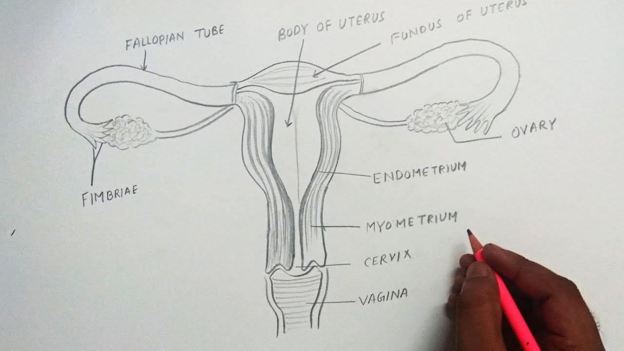
Female Reproductive Anatomy Drawing
Female Reproductive Anatomy Drawing - Drawing shows the uterus, myometrium (muscular outer layer of the uterus), endometrium (inner lining of the uterus),. Although a man is needed to reproduce, it is the woman who incubates the developing fetus and. Together they comprise the female reproductive system, supporting sexual and reproductive activities.the external genital organs, or vulva, are held by the female perineum.these are the mons pubis, labia majora and minora, clitoris, vestibule,. The vulva and its structures form the external genitalia. An incredibly distensible organ, the uterus can expand during pregnancy from around the size of a closed fist to become large enough to hold a full. The vagina is an elastic, muscular tube connecting the cervix of the uterus to the vulva and exterior of the body. The uterine tubes, the uterus, and the vagina. Web the female reproductive system is one of the most vital parts of the human reproductive process. Your vulva is the collective name for all your external genitals. The vagina, shown at the bottom of figure 27.9 and figure 27.10, is a muscular canal (approximately 10 cm long) that serves as the entrance to the reproductive tract.it also serves as the exit from the uterus during menses and childbirth. The ovaries produce eggs and hormones, and the fallopian tubes tran. Web the external female genitalia are a part of the female reproductive system, and include the: Together they comprise the female reproductive system, supporting sexual and reproductive activities.the external genital organs, or vulva, are held by the female perineum.these are the mons pubis, labia majora and minora, clitoris, vestibule,. Learn more about female reproductive organ anatomy. The function of your external genitals are to protect the internal parts from infection and allow sperm to enter your vagina. Web the uterus, also commonly known as the womb, is a hollow muscular organ of the female reproductive system that is responsible for the development of the embryo and fetus during pregnancy. Ovaries produce female sex hormones such as estrogen and progesterone as well as ova (commonly called eggs), the. Web a copulatory organ of the female reproductive system parts vaginal fornix (anterior/posterior/lateral parts), anterior and posterior walls of vagina, hymen, vaginal wall layers (mucosa [vaginal rugae → vaginal columns], lamina propria, muscular layer, adventitial layer). By teachmeseries ltd (2024) fig 1. Web the female reproductive anatomy includes both external and internal parts. Reproductive anatomy plays a role in sexual pleasure, getting pregnant, and breastfeeding.the urinary system helps rid the body of toxins through urination (peeing). Web female anatomy includes the external genitals, or the vulva, and the internal reproductive organs. Web the female reproductive anatomy includes both external and internal parts. Together they comprise the female reproductive system, supporting sexual and reproductive. Web the female reproductive system includes the ovaries, uterus, vagina, and vulva. The uterine tubes, the uterus, and the vagina. Web the uterus, also commonly known as the womb, is a hollow muscular organ of the female reproductive system that is responsible for the development of the embryo and fetus during pregnancy. An incredibly distensible organ, the uterus can expand. It is a distensible muscular tube which extends posterosuperiorly from the external vaginal orifice to the cervix. Ovaries produce female sex hormones such as estrogen and progesterone as well as ova (commonly called eggs), the. Anatomy and physiology of the female reproductive system. By teachmeseries ltd (2024) fig 1. Web the external female genitalia are a part of the female. The ovaries are a pair of small glands about the size and shape of almonds, located on the left and right sides of the pelvic body cavity lateral to the superior portion of the uterus. Web the uterus, also commonly known as the womb, is a hollow muscular organ of the female reproductive system that is responsible for the development. It houses the developing fetus during pregnancy. Web female anatomy includes the internal and external structures of the reproductive and urinary systems. Reproductive anatomy plays a role in sexual pleasure, getting pregnant, and breastfeeding.the urinary system helps rid the body of toxins through urination (peeing). The components of the external female genitalia occupy a large part of the female perineum. Web the uterus, also commonly known as the womb, is a hollow muscular organ of the female reproductive system that is responsible for the development of the embryo and fetus during pregnancy. This article looks at female body parts and their functions, and it provides an interactive diagram. A lot of people mistakenly use the term “vagina” to. Web the. This article looks at female body parts and their functions, and it provides an interactive diagram. The function of your external genitals are to protect the internal parts from infection and allow sperm to enter your vagina. This atlas is designed for medical students, residents and healthcare. By tim taylor last updated: The uterine tubes, the uterus, and the vagina. Web female anatomy includes the external genitals, or the vulva, and the internal reproductive organs. The outer walls of the anterior and posterior vagina are formed into longitudinal columns, or ridges, and the. Web reproductive system, female, anatomy: The components of the external female genitalia occupy a large part of the female perineum and collectively form what's known as the.. Web a copulatory organ of the female reproductive system parts vaginal fornix (anterior/posterior/lateral parts), anterior and posterior walls of vagina, hymen, vaginal wall layers (mucosa [vaginal rugae → vaginal columns], lamina propria, muscular layer, adventitial layer). This system of ducts connects to the ovaries, the primary reproductive organs. The uterus, or womb, is a hollow organ located centrally in the. Web the vagina is a muscular tube about 3 to 4 inches long.it is where a penis may enter during sexual intercourse. Web the female reproductive system is one of the most vital parts of the human reproductive process. The uterine tubes, the uterus, and the vagina. The function of your external genitals are to protect the internal parts from. This article looks at female body parts and their functions, and it provides an interactive diagram. Ovaries produce female sex hormones such as estrogen and progesterone as well as ova (commonly called eggs), the. Learn more about the anatomy and function of the female. Web the main parts of the female reproductive system include the following: The components of the external female genitalia occupy a large part of the female perineum and collectively form what's known as the. The ovaries are a pair of small glands about the size and shape of almonds, located on the left and right sides of the pelvic body cavity lateral to the superior portion of the uterus. It houses the developing fetus during pregnancy. It is a distensible muscular tube which extends posterosuperiorly from the external vaginal orifice to the cervix. Learn more about female reproductive organ anatomy. The vagina, shown at the bottom of figure 27.9 and figure 27.10, is a muscular canal (approximately 10 cm long) that serves as the entrance to the reproductive tract.it also serves as the exit from the uterus during menses and childbirth. Together they comprise the female reproductive system, supporting sexual and reproductive activities.the external genital organs, or vulva, are held by the female perineum.these are the mons pubis, labia majora and minora, clitoris, vestibule,. The vulva and its structures form the external genitalia. Web female reproductive system anatomy ovaries. Web learn about the anatomy of the female reproductive system including the vagina, uterus, fallopian tubes and ovaries with 3d interactive tutorials. Web reproductive system, female, anatomy: Web male reproductive system illustrations.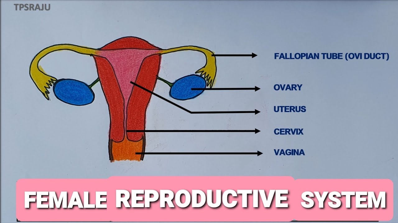
HOW TO DRAW FEMALE REPRODUCTIVE SYSTEM EASILY? THE STRUCTURE OF FEMALE
.jpg)
Female Reproductive System resource Imageshare
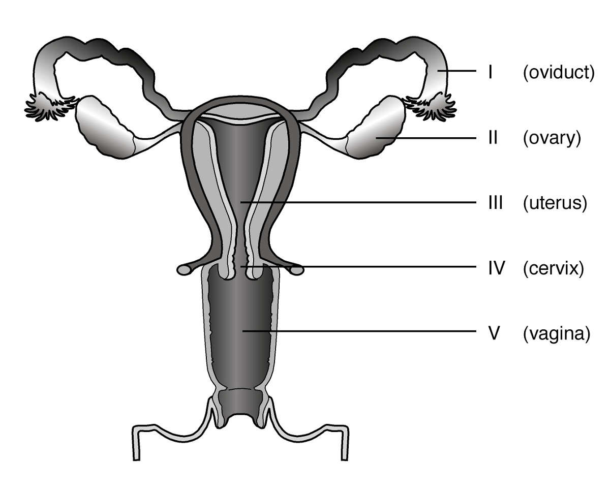
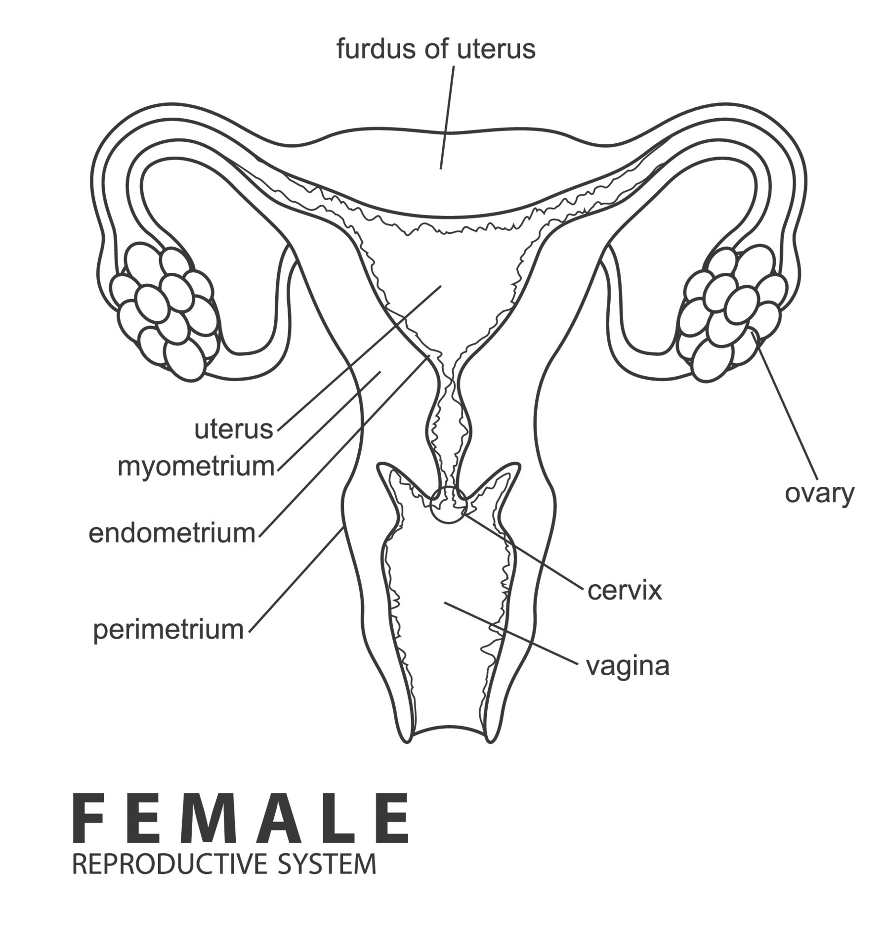
Diagrams of Female Reproductive System 101 Diagrams
Female Reproductive System Diagram Educative diagrams The Female
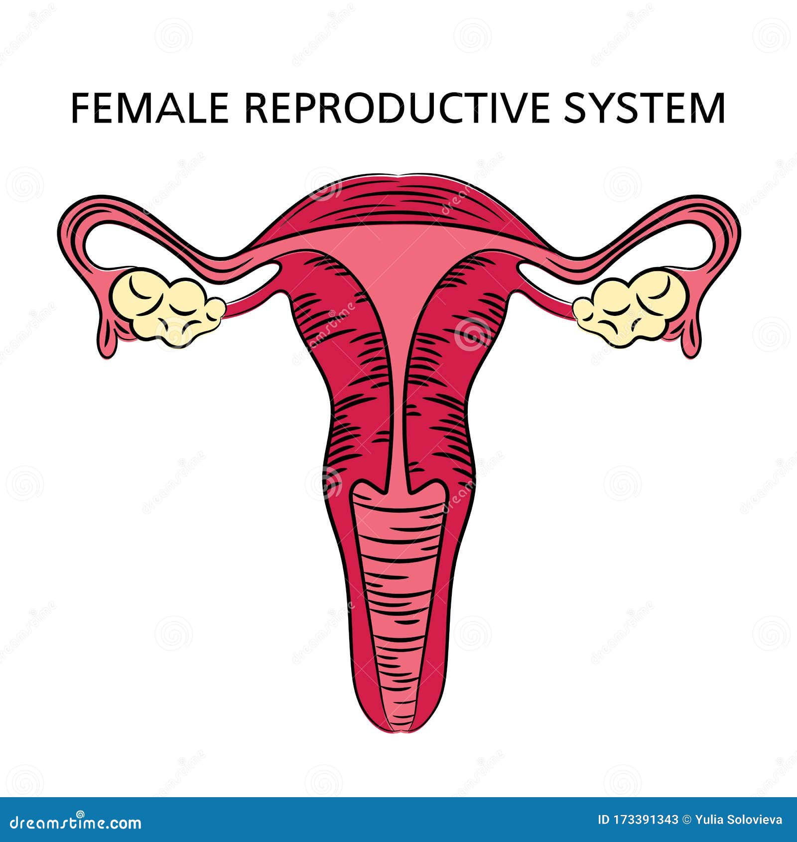
FEMALE REPRODUCTIVE SYSTEM Medicine Education Scheme Vector Stock

How to draw Female Reproductive system easily Step by step YouTube
How to draw female reproductive system easily step by step YouTube
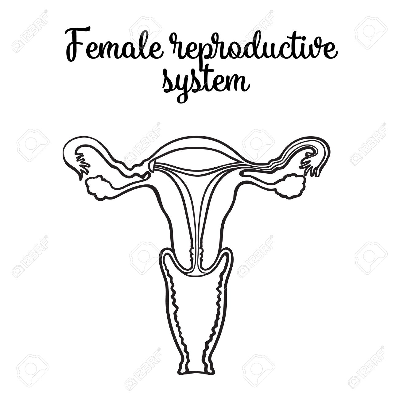
Female Reproductive System Drawing at GetDrawings Free download
Female reproductive system outline, Vector Illustration 22674066 Vector
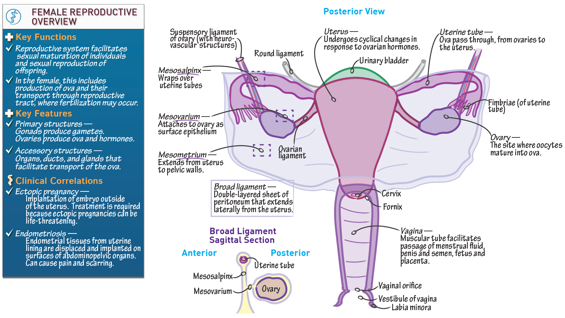
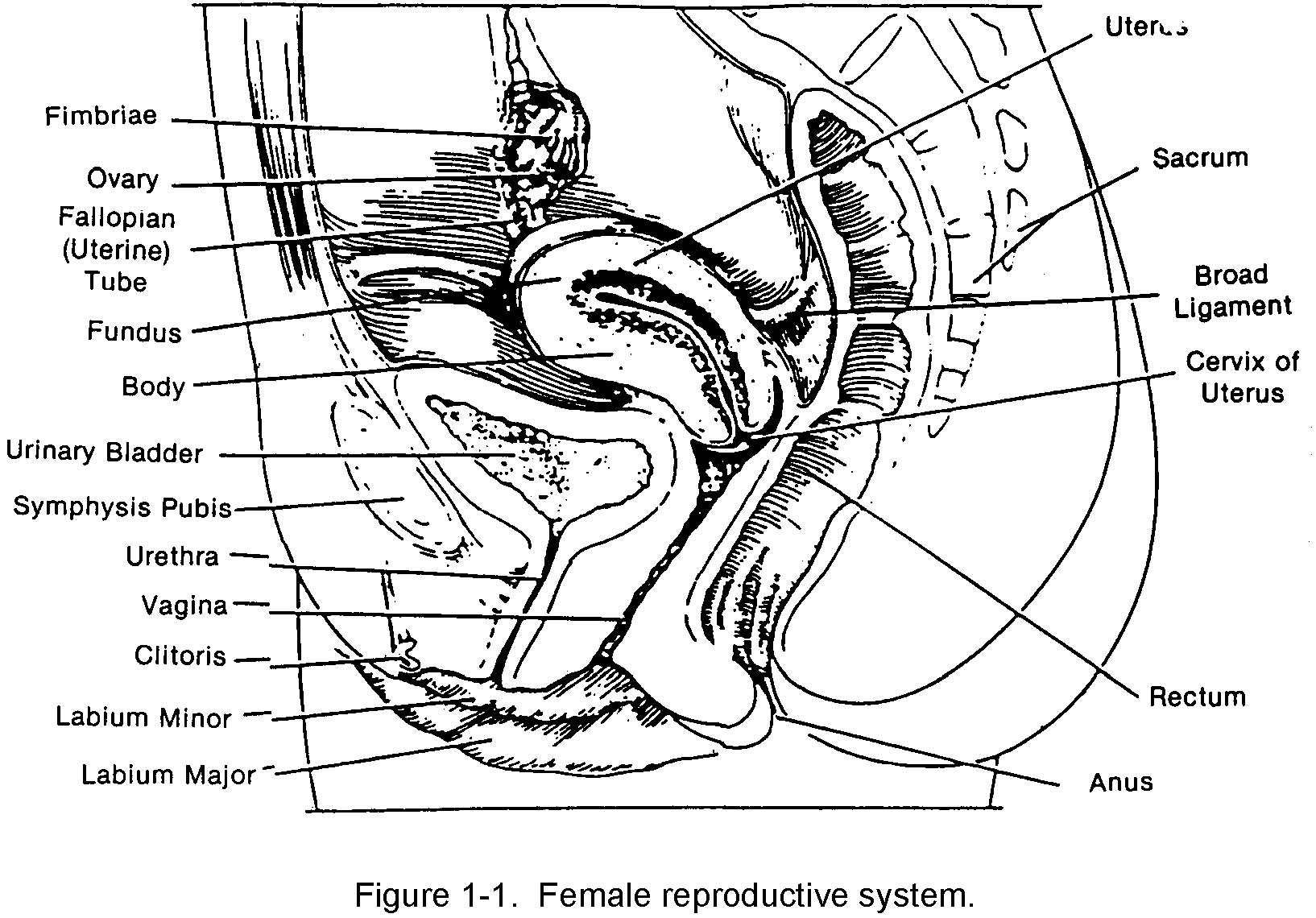
Anatomy & Physiology Anatomical Overview of the Female Reproductive
Although A Man Is Needed To Reproduce, It Is The Woman Who Incubates The Developing Fetus And.
The Function Of Your External Genitals Are To Protect The Internal Parts From Infection And Allow Sperm To Enter Your Vagina.
Web Learn About The Anatomy And Function Of The Vagina With Innerbody's 3D Illustrations.
Anatomy Of The Female Reproductive System;
Related Post: